London · 11–15 September 2026
Treeye at ESCRS 2026
The Optics and Vision Interest Group presented two posters at the 44th Congress of the ESCRS. If a printed code brought you here, everything is still on this page: the work, the tool behind it and how to reach us.
Who went
To meet people, mostly
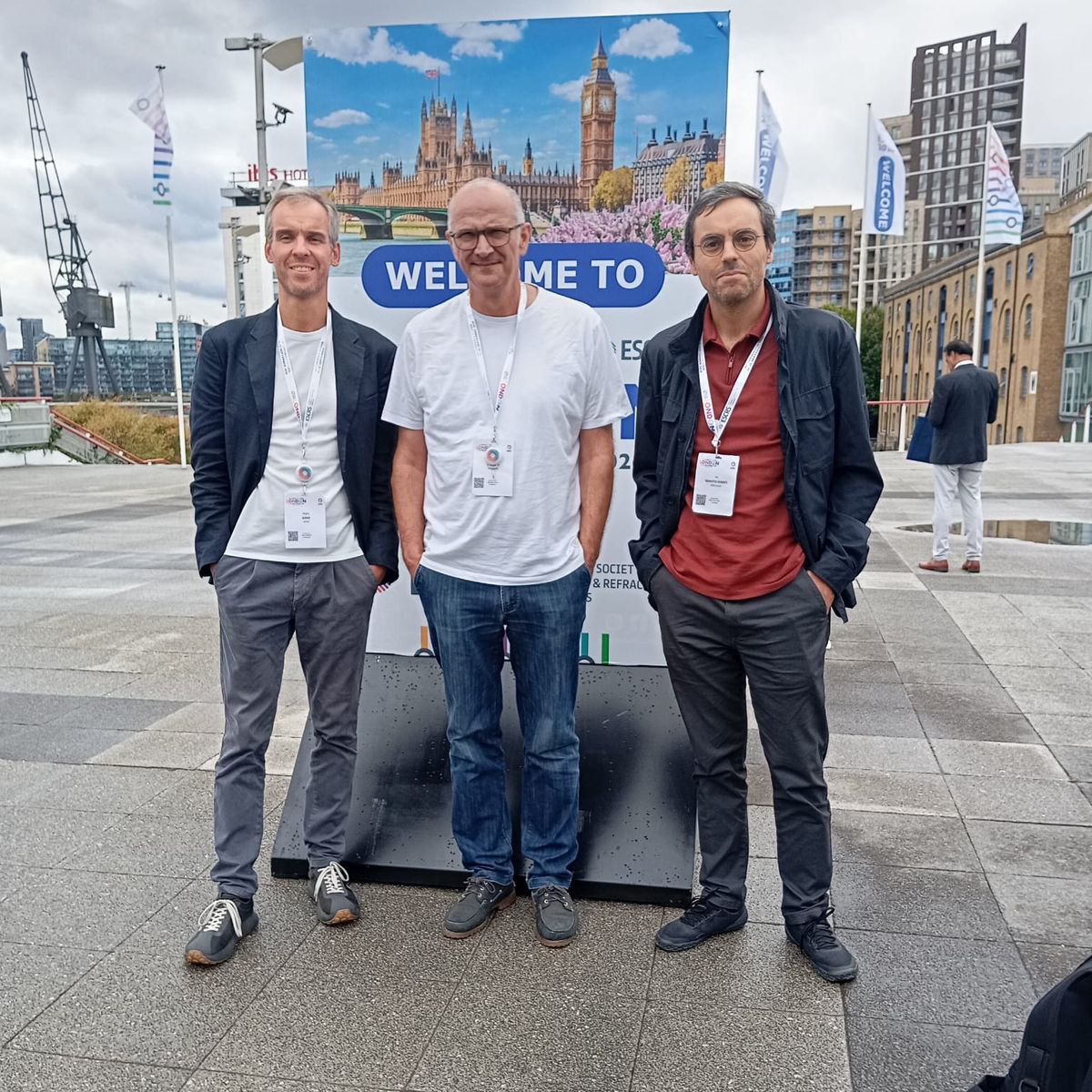
Pedro Serra, António Baptista and Ivo Soares went to London for the group. Treeye is an interest group rather than a company, and for us the point of a congress is the part that happens between sessions. We went to meet clinicians who’d use the tools, researchers with data worth pooling, and industry people who see where the instruments fall short.
What we presented
Seeing behind the iris
Anterior segment OCT is the reference standard for anterior segment biometry: fast, non-contact and highly accurate. But its reach stops at the iris. Light transmission through the iris is limited, and the little that returns is mixed with scattered light, so the structures immediately behind it, the ciliary sulcus among them, remain effectively invisible.
Our work presents an image-processing method that suppresses this non-anatomical signal and makes the structures of the retro-iris space visible in images already acquired in routine practice. We presented two posters: the first shows that the enhanced images allow repeatable sulcus-to-sulcus measurement, and the second tests the method against the clinical reference, ultrasound biomicroscopy, and finds good consistency between the two.
From this work we have built an image-processing and marking tool for anterior segment OCT. It isn’t released yet. There is a short demo on the tools page, and we would like to know whether it does what you would need it to.
-
Poster 4621 Sulcus-to-sulcus on AS-OCT
Sulcus-to-Sulcus Distance Measurement Using Anterior Segment Optical Coherence Tomography
Abstract
- Purpose
- To describe a multiscale spatial enhancement method for improving visualization of the retro-iridial space in anterior segment optical coherence tomography (AS-OCT) and to evaluate its clinical performance for sulcus-to-sulcus (STS) measurement.
- Setting
- Ophthalmology Clinic Vista Sánchez Trancón, Badajoz, Spain.
- Methods
- AS-OCT images acquired with the Anterion (Heidelberg) were processed using a multiscale spatial enhancement algorithm to reduce retro-iridial noise. Spatial decomposition at multiple scales enabled differentiation of anatomical structures from non-anatomical artefacts. Individual scale maps were combined into a consensus map reinforcing consistent anatomical features while suppressing noise. The enhanced images were used to measure STS distance with a dedicated digital caliper tool. In 30 eyes (30 patients), intra-rater repeatability was assessed by (1) repeated marking of the same image and (2) marking two separate images of the same eye. Agreement among white-to-white (WTW), spur-to-spur (SpTSp) and STS measurements was evaluated.
- Results
- Mean WTW, SpTSp and STS distances were 12.02 ± 0.35 mm, 12.11 ± 0.33 mm and 12.54 ± 0.54 mm respectively. STS measurements were larger than WTW (mean difference [MD] +0.50 ± 0.30 mm; 95% limits of agreement [LoA] −0.08 to +1.08 mm) and SpTSp (MD +0.41 ± 0.26 mm; 95% LoA −0.10 to +0.92 mm). WTW and SpTSp explained 51% and 61% of STS variability respectively. Intra-rater coefficients of repeatability were 0.07 mm (SpTSp) and 0.18 mm (STS) using the same image, and 0.14 mm (SpTSp) and 0.36 mm (STS) using different images.
- Conclusions
- Multiscale-enhanced AS-OCT imaging improves visualization of the retro-iridial space and enables repeatable STS measurements. This approach may offer a non-contact alternative for retro-iridial biometry, particularly in the context of phakic IOL sizing.
-
Poster 5370 AS-OCT against ultrasound
Sulcus-to-Sulcus Distance Comparison Measured Using Anterior Segment Optical Coherence Tomography and Ultrasound Biomicroscopy
Abstract
- Purpose
- To compare sulcus-to-sulcus (STS) distance measurements obtained using anterior segment optical coherence tomography (AS-OCT) and ultrasound biomicroscopy (UBM).
- Setting
- Instituto Salmantino de Oftalmología, Salamanca, Spain, and Ophthalmology Clinic Vista Sánchez Trancón, Badajoz, Spain.
- Methods
- Sixty eyes of 60 patients evaluated for phakic intraocular lens implantation were included. Imaging comprised AS-OCT (CASIA2, Tomey) and UBM (Quantel). UBM scans were obtained along the horizontal meridian by an experienced ophthalmologist, and the mean of three measurements was used as the reference STS value. AS-OCT B-scans were processed with a multiscale enhancement algorithm to improve retro-iridial visualization. STS (AS-OCT) was measured on enhanced images by a masked examiner using a custom caliper, while spur-to-spur (SpTSp) distance was obtained with the device caliper. Agreement was assessed using Bland–Altman analysis and intraclass correlation coefficients. The proportion of STS variability explained by SpTSp was calculated.
- Results
- Mean SpTSp, STS (AS-OCT) and STS (UBM) were 12.02 ± 0.36 mm, 12.39 ± 0.41 mm and 12.10 ± 0.37 mm respectively. AS-OCT STS measurements were larger than STS (UBM) by +0.23 ± 0.23 mm (95% limits of agreement −0.22 to +0.67 mm). The ICC for consistency was 0.83 (95% CI 0.73–0.90). After adjusting for systematic bias, 31%, 57% and 84% of eyes showed inter-device differences below 0.1, 0.2 and 0.3 mm respectively. SpTSp explained 69% (UBM) and 76% (AS-OCT) of STS variability.
- Conclusions
- Multiscale-enhanced AS-OCT enables reliable STS measurement with good consistency compared to UBM, but poor absolute agreement. Its non-contact nature and improved centration may offer practical advantages for retro-iridial biometry, particularly relevant for phakic posterior-chamber intraocular lens sizing.
Ivo Soares, Ángel Sánchez Trancón, António Baptista and Pedro Serra: Treeye, with the University of Beira Interior, the Physics Center of Minho and Porto Universities, Instituto Salmantino de Oftalmología and Ophthalmology Clinic Sánchez Trancón (Sanoptis).
For the record
At a glance
- Dates 11–15 September 2026
- Where ExCeL London
- Presented Posters 4621 and 5370
- There in person Pedro Serra, António Baptista and Ivo Soares
- The congress congress.escrs.org (opens in a new tab)
Questions about the posters or the tool? Write to us: [email protected]
While you’re here
The calculator is free to use
Our ICL Calculator is online and open source. It’s worth a look if you do Implantable Collamer Lens work.